Heart and Circulatory System
Stress Echocardiogram
Although FAA has historically relied upon stress ventriculography (radionuclide wall motion studies) for evaluating left ventricular function, techniques utilizing stress echocardiography are available that may augment or be substituted for radionuclide studies. The FAA has developed a protocol to assist airmen and their physicians in providing these reports:
- Echocardiographic imaging should be performed on current-generation two-dimensional equipment with digital capability using Microsonics, Freeland, or equivalent software providing a quad screen display and a closed cine loop. Doppler ultrasound and color flow techniques may provide additional useful information and should be included if available.
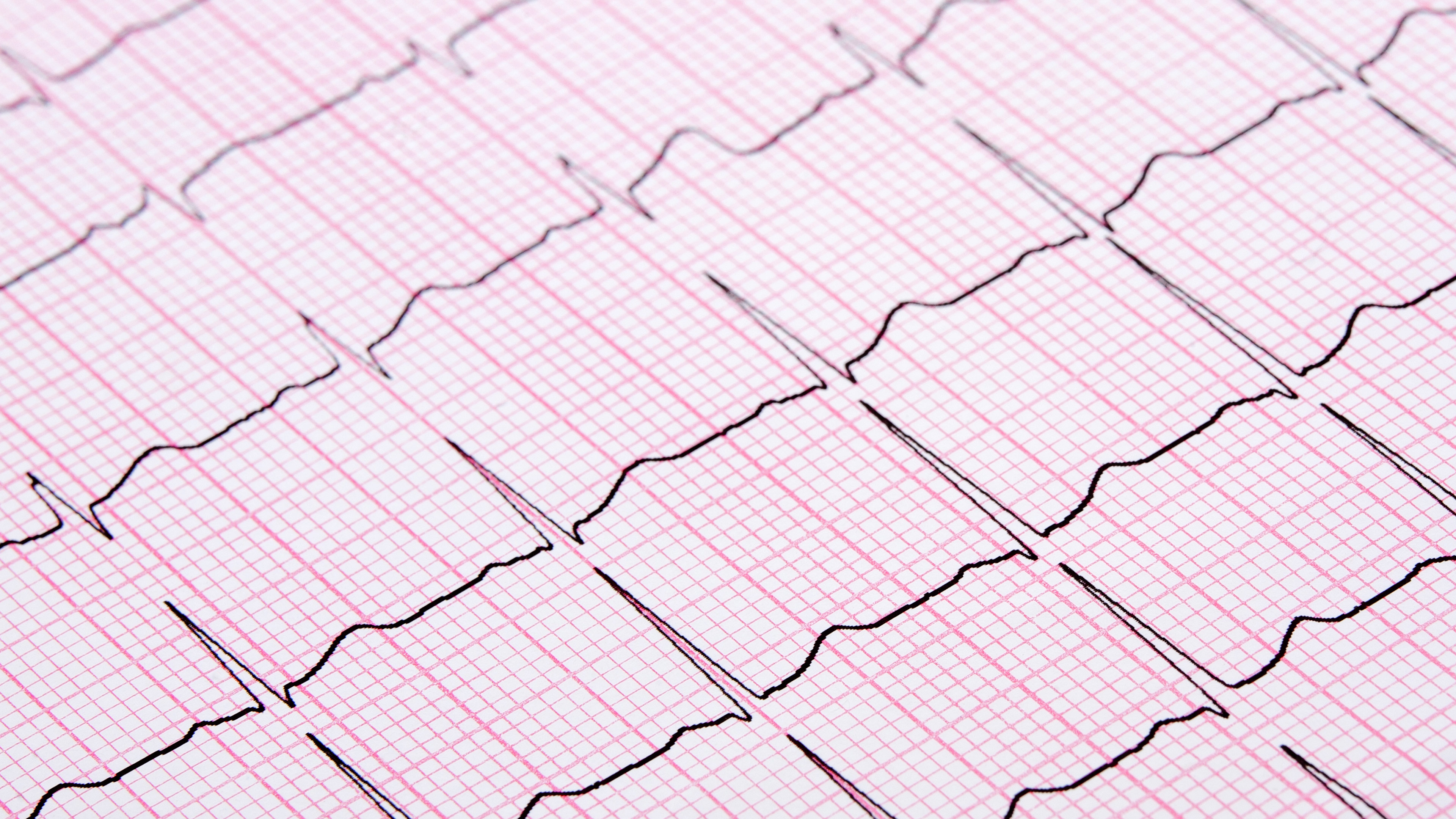
- All stress testing should achieve 100% of predicted maximum heart rate unless medically contraindicated or prevented either by symptoms or concurrent medication, such as beta adrenergic blockers, calcium channel blockers (especially diltiazem or verapamil), or digitalis preparations. These medications should be discontinued for at least 48 hours prior to testing in order to attain maximal stress and only after consulting with your attending physician. The blood pressure/pulse recordings at various stages and actual electrocardiographic tracings must be submitted. Tracings must include a rhythm strip. A full 12-lead ECG should be recorded supine and standing at rest, during hyperventilation while standing, one or more times during each stage of exercise, at the end of each stage, at peak exercise, and every minute during recovery for at least five minutes or until the tracings return to baseline. Computer-generated sample cycle electrocardiographic tracings are unacceptable in lieu of the standard tracings.
- Echocardiographic images should be obtained from four different views (usually parasternal long- and short-axis and apical four- and two-chamber views). The images should be acquired at rest, at each stage of exercise, and at peak exercise when utilizing supine or upright bicycle ergometer (a bicycle stress test). When exercising on a graded treadmill, imaging should be done at rest and within 90 seconds of completion of the test. For pharmacological testing, images must be acquired at rest, during low-dose stages, and following the maximum dose. Actual time of acquisition of the immediate post-exercise imaging should be noted.
- Atropine may be required in order to attain an adequate heart rate either during exercise or pharmacologic (dobutamine) testing.
- Additional testing modalities, including radionuclide scanning, may be required.
How/Where to Submit to the FAA
Helps you find the contact information for submitting your medical records.
Updated April 2016